Andrew Gore, O.D.
|
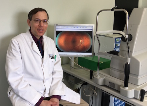
Dr. Gore grew up in Los Angeles. He received his undergraduate degree (in music and historical musicology) from UCLA, and began graduate school in musicology at UCLA when he was inspired by an eye exam to change his career goal to optometry. He then received the equivalent of a second bachelor's degree in biology and following this, attended graduate school at the Southern California College of Optometry, where he graduated in 1999. Because optometrists have gained so much responsibility with relation to ocular disease, Dr. Gore felt it necessary to pursue an optometry residency. (Only about 10% of U.S. optometry graduates complete residency training.)
|
The residency was a wonderful and exciting learning experience. It took place at the Standing Rock Indian Hospital Eye Clinic in Fort Yates, North Dakota, which is on a reservation 70 miles outside of Bismarck, North Dakota. During that one year on the reservation, Dr. Gore was exposed to a tremendous amount of ocular disease management in conjunction with a very experienced optometrist and an ophthalmologist. Upon his return to Southern California, Dr. Gore worked for a short time in a laser eye center and in corporate optometry and then accepted a position with ophthalmology groups in Santa Cruz.
Those four years in ophthalmology were also a time of great learning. During that time, Dr. Gore co-managed thousands of cataract surgery patients and learned to care for geriatric patients and specialty contact lens patients.
In September 2005, he purchased this Pasadena practice, which has been in the Citibank building since 1964. We are not sure exactly how old the practice is, but we know that the original founding doctor was licensed to practice in 1938!
Dr. Gore is looking forward to meeting you, helping you see better and taking care of your eye health.
2021 is our 57-year anniversary at this location!
It is amazing to contemplate this, but our founder, Dr. Gordon Lewis, moved into this office in 1964 when our building first opened. Our suite has been an optometry office ever since then. Dr. Gail Murphy took over in 1990, and then Dr. Gore became our doctor in 2005. During all of this time, we have done our best to provide patient, thorough, personalized eye care to our community, fighting against the trend of doctors' offices as factories. 2021 is also the 16-year anniversary of Dr. Gore's arrival at this location and his 22-year anniversary of becoming an optometrist.
OCT AND RETINAL CAMERA
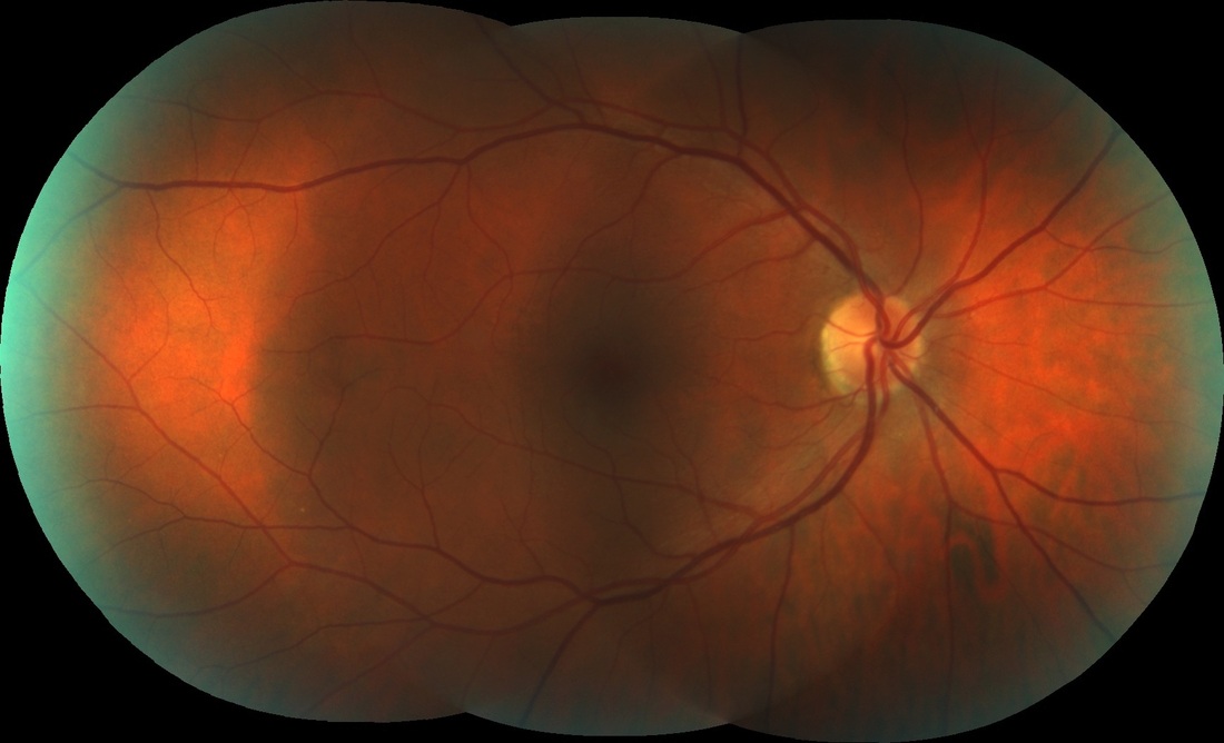
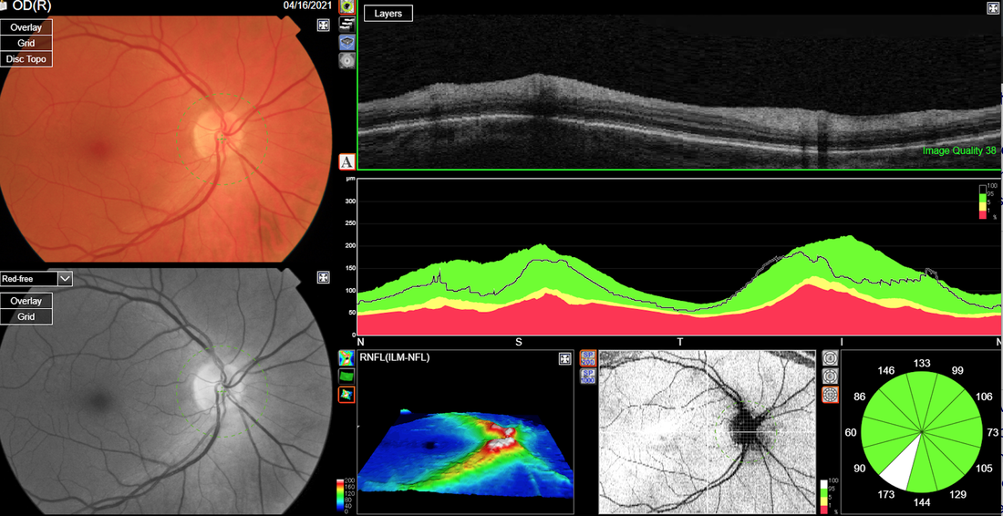
OUR RETINAL CAMERA AND OCT (optical coherence tomography) greatly enhance our ability to detect retinal problems. The retina is the inner surface of the eye and it is responsible for taking an image of what you see and converting it to an electro-chemical message that goes to your brain. Retinal imaging is not invasive and just takes a few minutes. In many cases, it is possible to take retinal images without dilating your eyes. The OCT device gives us a cross section of the layers of the retina, allowing us to see structures and damage inside the retina. The machine also measures and analyzes many of those structures with such fine detail that changes can be monitored over time. This miraculous machine has already revealed why for some of our patients, vision is decreased when everything else looks normal. Please note that retinal imaging is not a substitute for dilation and that it is often very important to do both dilation and retinal imaging. These are some reasons why retinal imaging is helpful:
- Screening images for all patients. When a doctor looks inside your eyes and scans around with a very bright light, it is possible to miss a problem, especially if it is a small problem. This is because (a) the doctor must work very quickly, as the light can be very painful to you, (b) patients tend to move around a lot, (c) the magnification of the eye's inner structures is very small, and (d) the doctor is human. A retinal image allows the doctor to take his time and look at a very magnified image of the retina, making it easier to detect a problem. We have discovered a countless number of people with ocular disease that may have been missed during normal exam procedures.
- Documenting anything unusual on the retina noticed during the exam (for example, unusual pigmentation, optic nerve structural changes, or diabetic changes). It is good to take a photograph so that we may compare the tissues as they change over time.
- Trying to decide if someone has glaucoma. Glaucoma is often a very tricky disease to diagnose. The optic nerve and nerve fibers are the parts that are affected by glaucoma, and they tend to assume a characteristic type of damage in glaucoma. However, there are many people who have funny looking optic nerves that are normal, and there are also people who have normal looking optic nerves that may have glaucoma just beginning. While we cannot put our trust in the OCT to give us a definite answer to this question, it often gives information that can help the doctor decide whether there is a problem and whether it is necessary to monitor the tissues in question to see if there are changes happening over time.

